He was kind of a handyman. He could fix anything.
![]()
Argonne National Lab Highlights: 1950-1959
1957
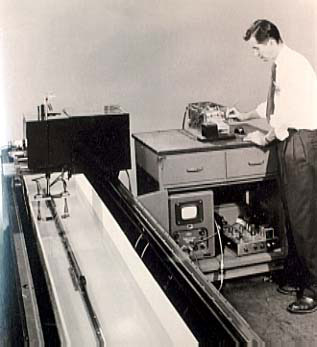
April 17 -- A short article by Argonne physicist W. Nelson Beck is received by The
Journal of the Acoustical Society of America. Printed in the July 1957 issue, it is the
first report of the discovery that ultrasound -- referred to as "an ultrasonic scanner and
recording system" -- has medical applications in making images of bones inside the
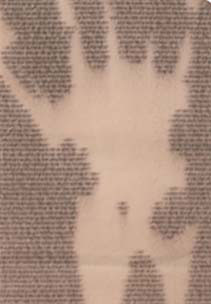
human body. The article is accompanied by history's first ultrasound image -- the
bones of Beck's hand and forearm. Beck had been working with ultrasound as a
non-destructive method for inspecting nuclear reactor fuel, when he was inspired to
put his arm inside the device and tune it to form an image of his bones.
"Taking Pictures with Sound"
Article appeared in Science, page 10, December 9, 1957.
Standard ultrasonic equipment is used, with two crystals - one to transmit, one to receive the vibrations. When one scanner encounters a flaw in the fuel element (or a bone in the hand), reception is interupted and a white space appears on the electrosensitive recording sheet (see photo).
Reprinted from The Journal of the Acoustical Society of America, Vol. 29, No. 7, 865, July, 1957
Copyright, 1957 by the Acoustical Society of America.
Ultrasonic Recording of the Bones in a Human Arm
W. N. BECK
Argonne National Laboratory, Lemont, Illinois
(Received April 17, 1957)
IN THE evaluation of an ultrasonic scanner and recording system, the effect of a transmission technique on human flesh at a frequency of 1 Mc was noted. By adjusting the sensitivity of the recording circuit it was possible to discriminate between flesh and bone. The figure on the left is a photograph of a recorded trace which indicates the location and the basic shape of bones in the hand and forearm. The two-dimensional figure is obtained by mechanically scanning an area with a small ultrasonic beam which transcribes a geometric "saw tooth". The recording is accomplished by a helix recorder which writes on electrosensitive paper. The scanner was developed primarily for the inspection of power reactor fuel elements, but this particular application might be of interest to other investigators.